


Past events at MIOT
For the 1st time in South India MIOT’s Experts Used an Advanced Stenting Technique to Successfully Treat a Life-Threatening Large Aneurysm at the Brainstem
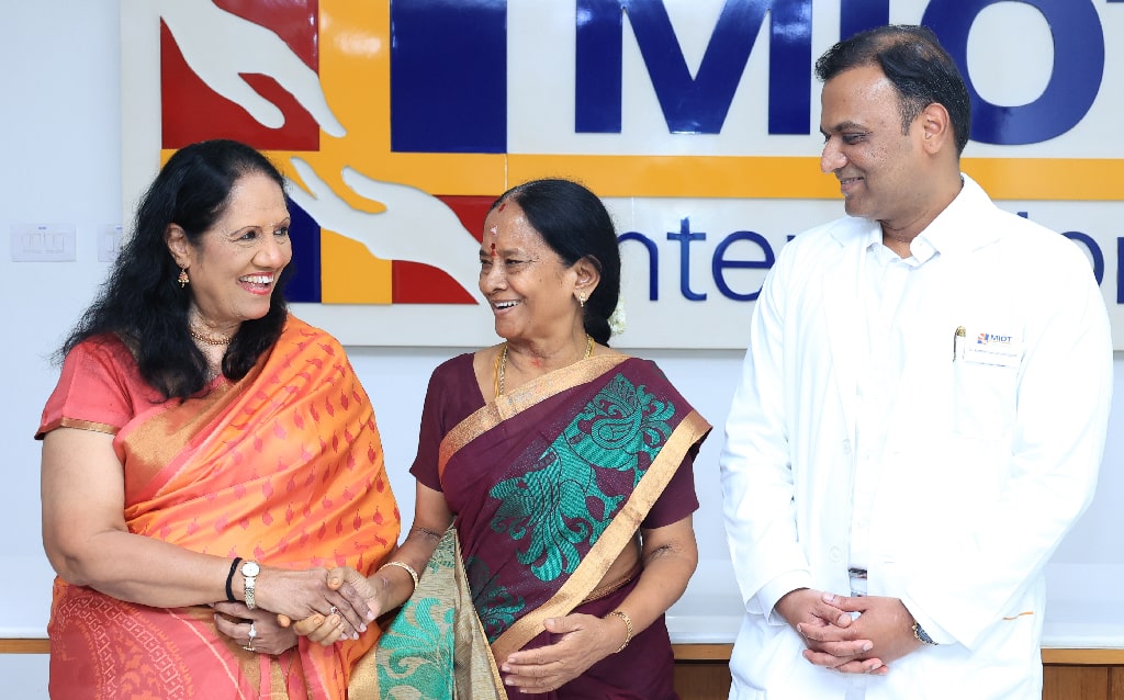
Photo seen from Left to Right: Mrs. Mallika Mohandas, Chairman, MIOT International, Mrs. Usha Rani, MIOT Patient who successfully underwent treatment for a 17mm wide necked aneurysm at the Brainstem, Dr. Karthikeyan Vanchilingam, Lead -Neuro Interventional Radiodiagnosis, MIOT International
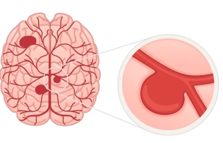
MIOT’s Interventional Neuroradiologists used an advanced stenting technique to successfully treat a life-threatening large aneurysm at the brainstem of a 65-year-old patient, without any complications.
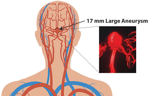
The 65-year-old Mrs. Usharani D, was diagnosed with an aneurysm in the artery which supplies blood to the brainstem called the basilar artery. It was 17 mm large, centrally located at the brain and was at risk of rupturing anytime, leading to sudden death. Additionally, the neck of the aneurysm was wide, measuring 11 mm, making it more complicated to treat.
Due to the size and complex location of this brain aneurysm in Mrs. Usharani, open surgery was impossible, and the surgical clipping method also involved high surgical complications.
Risks to the Patient
- At the risk of rupture anytime
- Potential for stroke
- High Surgical complications
- Impact of long-term blood thinners on her chronic kidney disease
- Risk of contrast dye affecting her renal function
- Anesthesia risks
- Recovery complications
For patients with incidental aneurysms that do not present with rupture, endovascular coiling is the treatment of choice. This minimally invasive (pinhole) procedure involves placing platinum wire coils within the aneurysm to block blood flow, thereby preventing rupture.
However, in Mrs. Usharani’s case, due to the wide neck of the aneurysm, the conventional treatment of endovascular coiling was not possible, as the coil could fall off from the aneurysm into the artery supplying blood to the brain, potentially causing a stroke.
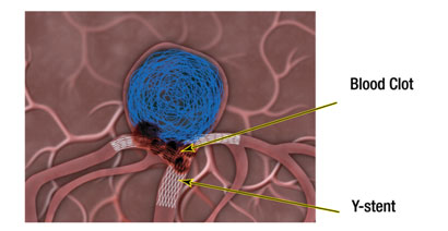
To prevent such coil displacement, Y-stent-assisted coiling was considered. This technique employs two stents placed in a Y-configuration holding the coil from displacement. However, this technique also poses a risk of stroke due to potential blood clotting at the Y-stent. To mitigate this risk, long-term blood-thinning medication is required, which could cause complications in the future due to her chronic kidney disease.
MIOT’s Innovative Treatment for Mrs. Usharani
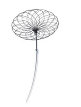
Considering the aneurysm’s large size, wide neck, complex location at the basilar artery, and the risks associated with Mrs. Usharani’s chronic kidney disease, MIOT’s Interventional Neuroradiologists, for the 1st Time in South India, used a Neqstent – a state-of-the-art device measuring 14 mm, the largest available, to treat her wide-necked aneurysm at the brainstem. This technique is not commonly used due to its need for specialised expertise. However, our Interventional Neuroradiologists with their expert skills successfully completed the pinhole procedure without any complications.
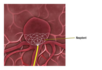
The Neqstent is designed like an inverted umbrella and can be placed at the neck of the aneurysm. In cases of wide-necked aneurysms, this stent helps prevent coil displacement. Additionally, this preserves the blood flow through normal arteries and significantly reduces the risk of stroke. Furthermore, this approach negates the need for long-term blood-thinning medications.
How was Neqstent-Assisted Coiling Performed?
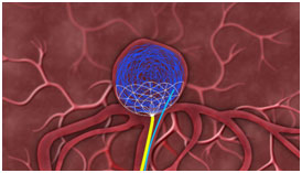
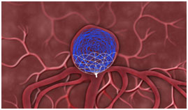
A small incision was made in Mrs. Usharani’s groin. Using the state-of-the-art Biplane CathLab with cone beam CT under X-ray guidance, a microcatheter over a guidewire was inserted into the artery and directed towards the aneurysm in the basilar artery of her brain. The Neqstent was inserted and unsheathed to cover the neck of the aneurysm. Once the deployment was confirmed to be satisfactory, the stability of the Neqstent device was checked by releasing any potential tension from the system. Subsequently, a catheter was navigated through the Neqstent into the aneurysm, and a fine platinum wire was coiled within the aneurysm until the desired packing density was achieved. This blocks the blood flow into the aneurysm. After completing the procedure, an electrolytic detachment system was used to detach the Neqstent device.
Over time, blood clots inside the coiled aneurysm, while also providing a scaffold to the coils. Eventually, the aneurysm would be sealed off from the circulation.
MIOT’s Interventional Neuro Suite
The intricate structure of the brain demands greater accuracy and speed to treat complicated aneurysms. Particularly, treating brain aneurysm located in the brainstem is highly challenging and requires cutting-edge technology, without which the treatment would be impossible.
For the first time in India, MIOT introduced the Biplane CathLab system with cone beam CT, 3D echo and software intelligence, all on a single platform. This system is supported by a 56″ LCD screen that visualizes Cone Beam CT images with high precision. The Biplane Neuro CathLab System provided clear 3D visualization of the brain and a real-time roadmap of target sites, allowing Interventional Neuroradiologists to navigate accurately. This enabled precise coiling and enhanced the safety and effectiveness of the treatment. By using this system, Interventional Neuroradiologists avoided multiple injections of contrast dye and repeated exposure to radiation, thereby preserving the patient’s renal function.
Mrs. Usharani’s Successful Recovery
Furthermore, Mrs. Usharani had another 6 mm aneurysm in the left tip of the major artery that supplies blood to the brain and eyes called the Internal Carotid Artery. She was also suffering from high blood pressure and chronic kidney disease and is under medication.
Considering her co-morbidities, another aneurysm in the internal carotid artery of her brain was also coiled during the same procedure. This approach avoided the need for additional angiograms and another major procedure under general anaesthesia. The patient’s condition was completely treated. Creatinine levels were kept stable throughout, ensuring her kidney function remained unaffected by the contrast dye used during the procedure.
By using the Neqstent instead of the Y-stent, the patient was successfully treated for this life-threatening condition without side effects or complications. She was spared from needing long-term blood-thinning medication and faced no future procedural restrictions. She has quickly regained her healthy, active life in a few days.